
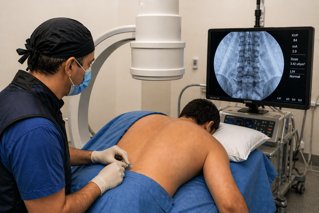
Did you ever wonder why an injection can completely relieve pain for a short period in one patient—yet provide little or no benefit in another?
The answer is rarely luck.
More often, it reflects whether the injection reached the true pain generator, or whether treatment was directed toward a structure that appeared abnormal but was not actually responsible for the symptoms.
At STAR Health, image-guided injections are not viewed simply as treatments for pain. They are often used first as diagnostic instruments within a systems-based evaluation, helping physicians verify which structure is generating symptoms before recommending additional procedures, surgery, or regenerative therapies.
Because when diagnosis improves, treatment decisions become more precise.
Pain Is a Signal—Not Simply a Structural Finding
One of the greatest misconceptions in musculoskeletal medicine is that pain always originates from the most abnormal finding on an MRI or X-ray.
Research consistently demonstrates that many imaging abnormalities occur in people who have no pain at all, including:
- Disc bulges and disc herniations
- Degenerative meniscal tears
- Rotator cuff degeneration
- Facet joint arthritis
- Tendon degeneration
These findings often represent age-related adaptations rather than the actual source of symptoms.
Pain develops when biology, mechanics, and nervous system signaling intersect—not simply because an image appears abnormal.
Within a systems-based model, every suspected pain generator remains a hypothesis until it is verified.
Diagnostic injections help answer one essential clinical question:
Is this tissue truly producing the patient’s pain—or is it simply present on imaging?
Diagnostic Injections Function as Clinical Hypothesis Testing
Every diagnostic injection represents a carefully designed clinical experiment.
Instead of assuming that a structure is responsible for pain, physicians temporarily alter the system and observe the response.
The process typically includes:
- Careful history and physical examination
- Correlation with imaging findings
- Image-guided placement of a small amount of local anesthetic
- Immediate reassessment of symptoms during functional movement
If pain substantially improves for the expected duration of the anesthetic, confidence increases that the targeted structure is contributing to symptoms.
If pain remains unchanged, the original hypothesis weakens, prompting clinicians to reconsider the diagnosis rather than escalating treatment unnecessarily.
Verification replaces assumption.
Historical Perspective — Localization Before Modern Imaging
Long before MRI scanners existed, physicians recognized that successful treatment depended on correctly identifying the source of pain.
Sir Charles Sherrington’s pioneering work in neurophysiology demonstrated that pain reflects complex neural signaling rather than tissue injury alone. His research helped shape our understanding that localization depends upon nervous system function as much as anatomy.
Centuries earlier, physicians practicing Ayurvedic medicine emphasized careful observation, functional examination, and therapeutic response when evaluating painful conditions—recognizing that symptoms often reflected disturbances across multiple body systems rather than isolated structures.
Different centuries.
Different technologies.
The same principle.
Diagnosis improves when hypotheses are tested rather than assumed.
Modern image-guided injections extend this philosophy by combining advanced imaging with real-time physiologic verification.
Brinjikji W, et al. Systematic Literature Review of Imaging Features of Spinal Degeneration in Asymptomatic Populations. American Journal of Neuroradiology.
Clinical Insight
An injection that temporarily changes a patient’s pain pattern often provides diagnostic information that imaging alone cannot reveal.
While imaging identifies anatomy, diagnostic injections help determine whether that anatomy is clinically meaningful.
Why Imaging Alone Can Be Misleading
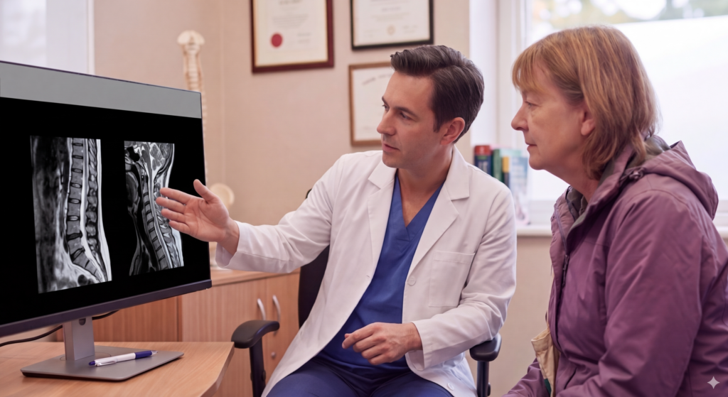
Imaging vs Function
MRI and CT imaging remain invaluable diagnostic tools.
However, they provide a static picture of anatomy.
Pain is dynamic.
Medical imaging can demonstrate:
- Structural integrity
- Degeneration
- Alignment
- Inflammation
- Tissue changes
It cannot directly demonstrate:
- Mechanical load transfer
- Dynamic movement dysfunction
- Central sensitization
- Neuromuscular compensation
- Functional pain reproduction
This helps explain why:
- Severe MRI abnormalities may produce little discomfort.
- Minimal imaging findings may correspond with significant disability.
Diagnostic injections restore a functional dimension to diagnosis by observing how symptoms change when a suspected pain generator is temporarily interrupted.
Precision Requires Navigation
Diagnostic accuracy depends upon precise placement.
At STAR Health, image guidance is selected according to the anatomy being evaluated.
This may include:
- Fluoroscopy for spinal procedures and selected joint injections
- Ultrasound for peripheral joints, tendons, bursae, muscles, and nerves
Navigation improves confidence that medication reaches the intended target while minimizing unintended spread.
Without image guidance:
- Medication distribution becomes less predictable.
- Diagnostic interpretation becomes less reliable.
- Clinical decision-making becomes more uncertain.
Precision supports better diagnosis—not simply more procedures.
How Diagnostic Injections May Help Reduce Unnecessary Surgery
When structural abnormalities are assumed to be responsible for symptoms without confirmation, patients may undergo procedures that ultimately fail to address the true pain generator.
Diagnostic injections may help clarify whether surgery is likely to address the patient’s symptoms by identifying which structures contribute to pain.
This additional diagnostic information can help avoid unnecessary escalation when symptoms arise from different tissues or from multiple interacting systems.
Examples include:
- Degenerative disc findings that are not producing symptoms
- Meniscal abnormalities unrelated to knee pain
- Rotator cuff degeneration that is not the primary pain source
- Facet degeneration identified incidentally on imaging
By improving diagnostic confidence, physicians can better determine when conservative management, rehabilitation, interventional procedures, regenerative medicine, or surgical consultation may be appropriate.
Local Care, Global Science
Patients throughout Fort Wayne and Northeast Indiana frequently search for answers before committing to injections or surgery.
Common questions include:
- Diagnostic nerve block
- Image-guided injections near me
- Interventional pain specialist Fort Wayne
- How to avoid unnecessary spine surgery
- Image-guided joint injections
At STAR Health, current evidence in musculoskeletal medicine is integrated with physician-led clinical reasoning to help determine whether an injection should be used to clarify diagnosis, provide symptom relief, or both.
When Diagnostic Injections Become Therapeutic
Once a pain generator has been reasonably confirmed, treatment options become more focused.
Depending on the diagnosis and the patient’s goals, management may include:
- Targeted anti-inflammatory injections
- Progressive rehabilitation
- Activity modification
- Biomechanical optimization
- Regenerative medicine when clinically appropriate
- Ongoing reassessment of functional recovery
The intervention follows the diagnosis.
Not the other way around.
Did You Know?
Numerous imaging studies have shown that disc bulges, tendon degeneration, meniscal tears, and other musculoskeletal changes are frequently found in individuals without pain. This is one reason physicians often combine imaging with physical examination and, when appropriate, diagnostic injections before determining the source of symptoms.
Author Expertise (E-E-A-T)
Care at STAR Health is led by Dr. Joseph Fortin, DO, a board-certified physician in Physical Medicine & Rehabilitation with subspecialty certification in Interventional Pain Management.
His diagnostic philosophy emphasizes careful clinical reasoning, image-guided precision, and verification before intervention—helping patients better understand the source of pain before considering more invasive treatment options.
Reader Reflection
Have you ever been advised to undergo an injection or surgery based primarily on an MRI or X-ray?

Understanding whether that finding truly matches your symptoms may be one of the most important steps before deciding on the next stage of treatment.
Frequently Asked Questions
Are diagnostic injections the same as steroid injections?
No. A diagnostic injection often uses a small amount of local anesthetic to determine whether a structure is causing pain. Therapeutic injections may include medications intended to reduce inflammation or support symptom management.
Will a diagnostic injection permanently relieve pain?
Its primary purpose is diagnostic. Temporary symptom improvement may help physicians identify the source of pain, but lasting treatment depends on the underlying diagnosis.
Why is image guidance important?
Image guidance helps improve placement accuracy, increasing confidence that medication reaches the intended structure while reducing diagnostic uncertainty.
Can an MRI identify the exact source of pain?
Not always. Imaging provides valuable anatomical information but must be interpreted alongside history, physical examination, and, in some cases, diagnostic injections.
When should diagnostic injections be considered?
They may be appropriate when physical examination and imaging suggest multiple possible pain generators or when additional confirmation may help guide treatment decisions.
Before moving toward more invasive treatment, make sure the diagnosis is as precise as possible.
STAR Health — Fort Wayne, Indiana
Physician-led, image-guided evaluation focused on verification before intervention.
Confirm the source. Guide the treatment. Make decisions with greater confidence.