
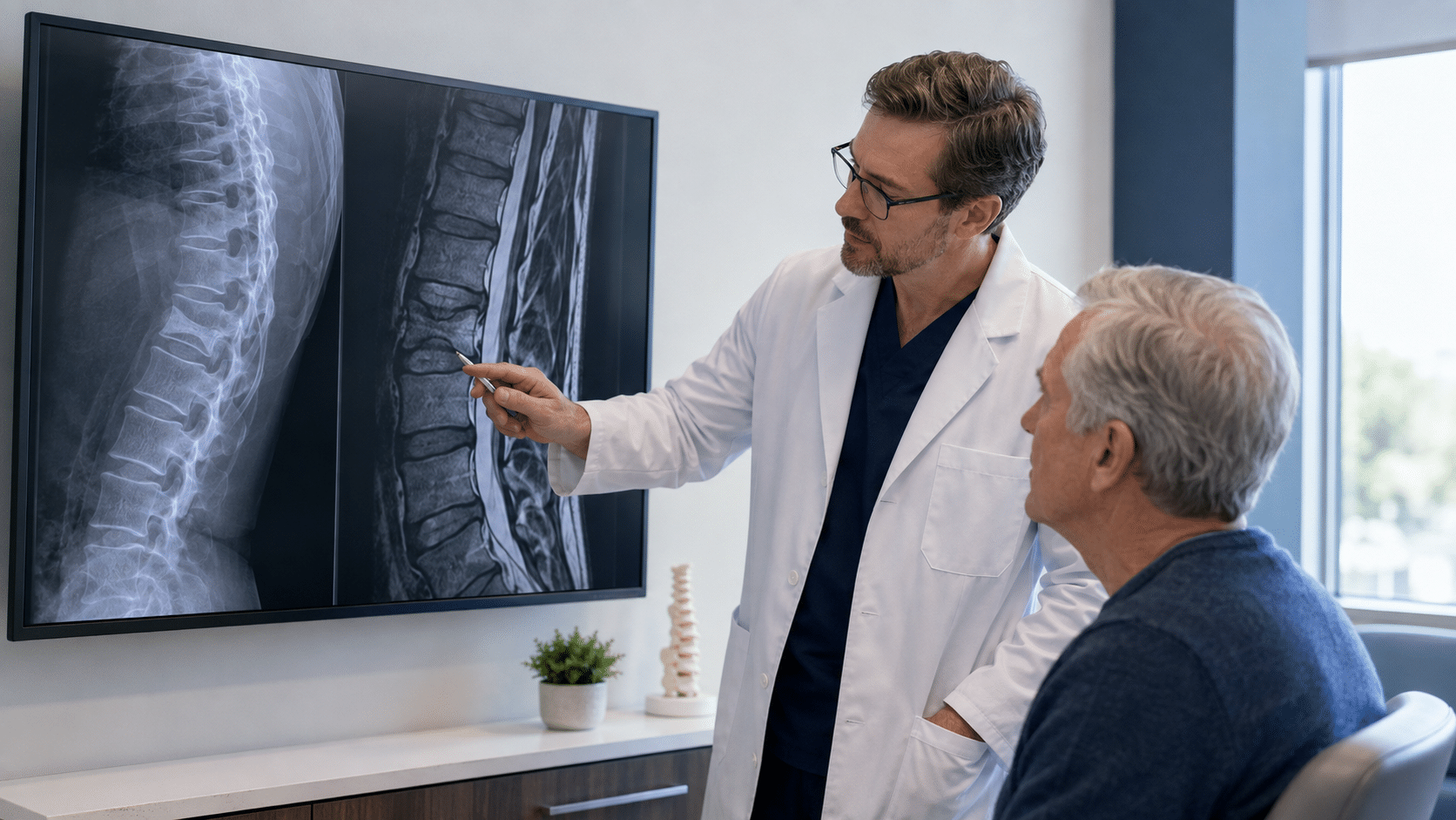
The first clue may be sharp pain after bending, coughing, or lifting. No dramatic fall—only a spine that suddenly refuses to carry the body as it did yesterday.
The danger can spread. A vertebral compression fracture may alter posture, restrict breathing, discourage walking, and erode independence.
Kyphoplasty for compression fractures is often described as a pain procedure. In selected patients, it is vertebral stabilization. Yet not every fracture requires it. The diagnostic question comes first: Is this vertebra still an active source of pain and instability, and would stabilization offer more benefit than conservative care?
The Fracture Changes a System, Not Just a Bone
The spine is a living load-bearing column. Bone, discs, joints, ligaments, muscles, and nerves coordinate movement and balance.
When one vertebral body loses height, three connected systems can change:
- Structure: Wedge-shaped collapse can shift spinal alignment.
- Signaling: Fracture-site inflammation and micromotion can activate pain pathways during standing or turning.
- Load: Force is redistributed while muscles work harder to keep the body upright.
The body guards. Stride shortens. Sitting replaces walking. Over time, protection may reduce strength, respiratory excursion, balance, and confidence. The fracture is local; its consequences can become systemic.
Diagnosis Before Stabilization
A compressed vertebra on X-ray does not automatically identify today’s pain source. Some deformities are healed; others are new and mechanically painful. Some coexist with disc, nerve, infection, or tumor-related disease.
Evaluation may integrate:
- Symptom timing and behavior
- Focal tenderness and movement-provoked pain
- Neurologic examination
- X-ray, MRI, or CT findings
- Bone marrow edema or another sign of fracture activity
- Bone quality and future fracture risk
The RSNA and ACR patient guide emphasizes evaluation and imaging before vertebral augmentation. STAR Health can connect this workup with musculoskeletal and orthopedic medicine and pain management when another structure may contribute.
The goal is not to find a reason to perform kyphoplasty. It is to determine whether the fracture explains the system failure.
Clinical Visualization: When “Rest” Becomes a Trap
Imagine an older adult who feels abrupt mid-back pain while reaching into a cabinet. A brace makes lying down tolerable, but standing remains sharply painful.
Weeks pass. The patient walks less, sleeps in a recliner, and avoids cooking because standing hurts. The legs weaken. Breathing feels shallower. One fracture now affects endurance and independence.
If examination and updated imaging confirm an active, pain-producing fracture, stabilization may create an opportunity to move again. If the fracture is healed or pain comes from elsewhere, kyphoplasty may add risk without solving the problem.
What Kyphoplasty for Compression Fractures Actually Changes
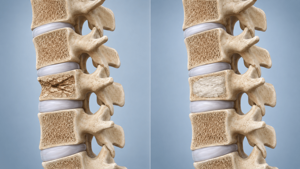
Under imaging guidance, a channel is advanced into the fractured vertebral body. An inflatable bone tamp creates a cavity and may partially restore height when anatomy allows. The balloon is removed, and bone cement is placed to stabilize the vertebra internally.
Kyphoplasty does not require rods, plates, or pedicle-screw instrumentation. The cement remains in the vertebra, however, and changes local material properties. Cement volume, distribution, viscosity, timing, and imaging control therefore matter.
The main objective is not cosmetic straightening. It is to reduce painful fracture motion and improve load tolerance. Results vary, and kyphoplasty does not treat the osteoporosis or other disease that weakened the bone.
Clinical Insight: Stabilization Is a Timing Decision
Clinical Insight: The meaningful comparison is not “procedure versus no procedure” in the abstract. It is the risk of intervening compared with the risk of allowing a confirmed, active fracture to continue limiting mobility.
Medication, activity modification, bracing, and guided rehabilitation are appropriate for many patients. When severe concordant pain persists, immobility has consequences of its own. Fracture activity, health status, symptoms, and functional trajectory—not the calendar alone—should guide the decision.
Survival and Function: What the Evidence Can—and Cannot—Say
Compression fractures are associated with reduced mobility, loss of independence, and increased mortality, particularly among older adults with osteoporosis and other illnesses.
A 2020 meta-analysis of more than two million patients found vertebral augmentation was associated with a 22% lower mortality risk than nonsurgical management over follow-up as long as ten years. This does not prove the procedure lengthens life; much evidence was observational, and patient selection may affect the result. Review the specific Radiology meta-analysis.
The responsible conclusion is narrower: a vertebral fracture can be both a marker and a driver of systemic decline, and restoring mobility may matter beyond pain. Kyphoplasty should never be presented as a survival guarantee.
Why Technique Matters: Unipedicular Precision
Dr. Joseph Fortin typically uses a unipedicular approach when anatomy permits. One access path may reduce tissue disruption and procedural or fluoroscopy time.
But unilateral access is not automatically better. Cement must still reach a safe, effective distribution. If anatomy or fracture morphology requires bilateral access, the approach should change. Precision means adapting technique to the vertebra.
Experience, Expertise, Authority, and Trust
STAR Health’s kyphoplasty care is directed by Joseph D. Fortin, DO, a board-certified Physical Medicine and Rehabilitation physician with subspecialty certification in Interventional Pain Management and certification in Clinical Densitometry.
Vertebral fracture care requires more than procedural access. It requires integrated interpretation of imaging, pain behavior, neurologic status, skeletal health, biomechanics, and recovery capacity. Every recommendation begins with diagnosis.
Did You Know?
A compression deformity can remain visible after the fracture has healed. MRI marrow edema can help identify fracture activity, while the examination tests whether that level matches the patient’s pain.
“A fracture appears on X-ray” and “this fracture should be stabilized” are not the same conclusion.
Historical Perspective: From Vertebroplasty to Kyphoplasty
Modern vertebral augmentation began in Amiens, France, where Jacques Galibert and Hervé Deramond performed vertebroplasty in 1984 for a painful vertebral hemangioma. Their report followed in 1987.
Kyphoplasty later added an inflatable bone tamp before cement placement. The technology changed; the central idea remained: reinforce weakened bone through a small access route while limiting disruption to surrounding tissue.
Local Care, Global Science
People seeking kyphoplasty for compression fractures in Fort Wayne may have a recent osteoporosis-related fracture, persistent pain after bracing, postural change, or uncertainty about an old deformity.
STAR Health applies global imaging, biomechanics, augmentation, and rehabilitation principles in Northeast Indiana. The objective is to identify the pain generator, choose an appropriate strategy, and plan the bone health and movement recovery that follow.
Frequently Asked Questions
Is kyphoplasty for compression fractures appropriate for everyone?
No. Many improve conservatively. Kyphoplasty may be considered when an active fracture is confirmed, symptoms match, and expected benefit outweighs risk.
How is an active fracture identified?
History, examination, X-ray, MRI, or CT may be used. MRI marrow edema can show activity, but imaging must match the clinical pattern.
Can kyphoplasty restore vertebral height?
It may partially restore height in some mobile, relatively recent fractures. Restoration is not guaranteed; stabilization is the primary objective.
Does it prevent future compression fractures?
No. Kyphoplasty treats a selected fracture; it does not cure osteoporosis. Bone-health treatment, fall-risk reduction, nutrition, and progressive loading remain important.
What are the risks?
Risks include bleeding, infection, cement leakage, medication or anesthesia reactions, nerve injury, embolic complications, persistent pain, and additional fractures.
What is recovery like?
Many procedures are outpatient, but recovery varies. Activity, wound care, medication, and rehabilitation instructions should follow the treating physician’s plan.
If a compression fracture limits standing, walking, breathing, or confidence, the next step is not automatically a procedure. It is a precise diagnosis.
STAR Health evaluates whether the fracture is active, whether it explains the symptoms, and whether stabilization fits the larger recovery plan.
📍 STAR Health — Fort Wayne, Indiana
📞 260-459-7313
🕒 Physician-directed, time-rich evaluation
Understand the fracture. Stabilize when appropriate. Rebuild the system.
Contact us to schedule a consultation today!